 what’s app : +91 9290097068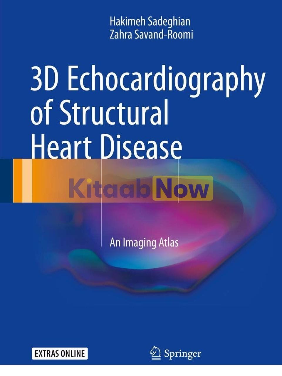
-22%
3D Echocardiography of Structural Heart Disease: an Imaging Atlas
Generic
$ 98.55
Call 1800-313-2353 for expert assistance!
- Questions & Answers
DescriptionThis atlas presents outstanding three-dimensional (3D) echocardiographic images of structural heart diseases, including congenital and valvular diseases and cardiac masses and tumors. The aim is to enable the reader to derive maximum diagnostic and treatment benefit from the modality through optimal image acquisition and interpretation. To this end a wide range of instructive individual cases are depicted, with sequential arrangement of all images and views of diagnostic value, including 3D zoom, full-volume, and live 3D images. For each case, a key lesson is highlighted and attention is drawn to aspects of relevance to diagnosis and treatment. In addition, readers will have online access to echocardiographic video clips for each patient. The closing part of the book examines the role of 3D echocardiography in structural heart disease interventions. The superb quality of the illustrations and the range of cases considered ensure that this atlas will be an excellent visual learning tool and an ideal aid for cardiology residents and fellows in day-to-day clinical practice.Table of Contents1. Chapter 1 Degenerative Mitral Valve Disease 2. Chapter 2 Rheumatic Mitral Stenosis 3. Chapter 3 Aortic Valve Disease 4. Chapter 4 Tricuspid Valve Disease 5. Chapter 5 Pulmonary Valve Disease 6. Chapter 6 Malfunction and Other Complications After Heart Valve Surgery 7. Chapter 7 Paravalvular Leak of Prosthetic Valves 8. Chapter 8 Infective Endocarditis (IE) 9. Chapter 9 ASDs and PFO 10. Chapter 10 VSD, PDA, Coarctation of Aorta, Subvalvular AS 11. Cardiac Mass 12. Intervention in Structural Heart Disease 13. Degenerative Mitral Valve Disease 14. Introduction 15. Case 1: Fibroelastic Deficiency, Prolapse of A2, Moderate Mitral Regurgitation (MR) 16. Case 2: Fibroelastic Deficiency (FED ), Severe Mitral Regurgitation, Prolapse and Rupture of 17. Case 3: Fibroelastic Deficiency (FED ), Rupture of Chorda of A2, Severe MR 18. Case 4: Forme Fruste, Rupture of Chorda of P2, Severe MR 19. Case 5: Rupture of Chorda of P2, Severe MR, Barlow Disease 20. Case 6: Barlow Disease, Rupture of Chorda of P2, Severe MR 21. Case 7: Barlow Disease, Severe Mitral Regurgitation, ¬Elongation of Chorda of P2 22. Case 8: Barlow Disease, Rupture of Chorda of Lateral Side of P2 (Between P1 and P2), Prola 23. Case 9: Barlow Disease, P2 Rupture, Severe MR 24. Case 10: Barlow Disease, Severe Mitral Regurgitation, and Rupture of Chorda of A2 and A3 25. Case 11: Severe Functional and Organic MR Due to Prolapse of A2 in a Post CABG Patient ( 26. Case 12: Severe MR Due to Fibroelastic Deficiency and Flail of A2 27. Case 13: Severe MR Due to Barlow Disease 28. Case 14: Severe Mitral Regurgitation Due to Flail of P3 (Forme Fruste) 29. Case 15: Severe MR Due to Flail P2 (FED ) 30. Case 16: Severe MR Due to Flail of P2 (FED ) 31. References 32. Rheumatic Mitral Stenosis 33. Introduction 34. Case 1: Severe Mitral Stenosis, Mild MR, Mild AS, Moderate AI, No LA, and LAA Clot Suitable fo 35. Case 2: Severe MS, No MR, Mild AS, Mild to Moderate AI, and LAA Clot 36. Case 3: Severe MS, Mild MR, Mild AI, No LAA, and LAA Clot Suitable for PTMC 37. Case 4: Severe MS, Severe Spontaneous Contrast in LA, and LAA Clot 38. Case 5: Severe MS, Severe MR, Severe TR, and Severe Spontaneous Contrast in LA 39. Case 6: Severe MS, Moderate MR, and a Mobile Mass on Ventricular Side of Aortic Valve 40. Case 7: Severe MS, Mild MR, No LA, and LAA Clot 41. Case 8: Severe MS, Mild MR, Moderate AS, Moderate AI, No LA, and LAA Clot 42. Case 9: Severe MS, Mild MR, Moderate AI, No LA, and LAA Clot 43. Case 10: Mild Mitral Stenosis due to Severe Mitral Annulus Calcification and Senile Degener 44. Case 11: Severe Mitral Stenosis, Severe Mitral Regurgitation, Severe Tricuspid Regurgitation, 45. Case 12: Severe Mitral Stenosis, Mild Mitral Regurgitation, Severe Tricuspid Regurgitation, an 46. References 47. Aortic Valve Disease 48. Aortic Stenosis 49. Definition 50. How to Approach 51. Aortic Regurgitation 52. Case 1: Severe Aortic Regurgitation 53. Case 2: Critical AS, Severe AI 54. Case 3: Moderate Valvular AS, Bicuspid Aortic Valve, and Interrupted Aortic Arch 55. Case 4: Bicuspid Aortic Valve with Moderate Valvular Aortic Stenosis 56. Case 5: Bicuspid Aortic Valve, Mild AS, Mild AI 57. Case 6: Low Flow, Low Gradient Severe AS 58. Case 7: Paradoxical Low Gradient Severe AS 59. Case 8: Severe AS Post-CABG 60. Case 9: Bicuspid Aortic Valve with Severe AS and Severe AI 61. Case 10: TAVI Procedure 62. Case 11: Visualization of Four Pulmonary Veins and Measurement of Aortic Annulus for TAVI 63. Case 12: Type A Aortic Dissection 64. References 65. Tricuspid Valve Disease 66. Tricuspid Stenosis, Tricuspid Regurgitation 67. Case 1: Severe TS, Severe TR, Severe MS, Mild MR 68. Case 2: Severe TS Post-MVR 69. Case 3: Severe TS and Moderate TR and Severe MS 70. Case 4: Severe TS and Severe TR, Severe MS, Mild MR 71. Case 5: Carcinoid Disease with Severe TR and Severe PI 72. What is a Carcinoid Tumor? 73. What is a Carcinoid Heart? 74. What is the Cause of Involvement of the Right-Sided Valve in a Carcinoid Heart? 75. How is 5-HIAA Used? 76. Is There Anything Else that Affects 5-HIAA Level? 77. Case 6: Severe TR Due to Quadricuspid Tricuspid Valve 78. What is the Congenital Anomaly of Tricuspid Valve? 79. Tricuspid Atresia 80. Congenital Tricuspid Stenosis 81. Congenital Cleft of the Anterior Leaflet 82. What is the Variation in Number of Cusps in the Tricuspid Valve? 83. Case 7: Severe TS and Severe TR Due to Rheumatismal Involvement 84. When Do You Send Patient for Right-Sided Valve Surgery Along with a Good Left-Sided Prost 85. References 86. Pulmonary Valve Disease 87. Pulmonary Stenosis, Pulmonary Regurgitation 88. Case 1: Severe Valvular and Subvalvular Pulmonary Stenosis, Bicuspid Pulmonary Valve 89. Case 2: Moderate Valvular Pulmonary Stenosis with Partially Aneurysmal IAS and PFO and Small 90. Case 3: Moderate Valvular Pulmonary Stenosis with Right Ventricular Systolic Dysfunction 91. Case 4: Severe PI Due to Infective Endocarditis 92. Case 5: Bicuspid Pulmonic Valve with Mild PS 93. References 94. Malfunction and Other Complications After Heart Valve ¬Surgery 95. Case 1: MVR with Bileaflet Prosthetic Mitral Valve and TVR with Bioprosthetic 96. Case 2: Valve in Ring for TV 97. Case 3: Severe TR on TV Ring Annuloplasty 98. Case 4: MVR and AVR and TV Ring Annuloplasty with Moderate TR 99. Case 5: Incomplete MV Ring Annuloplasty 100. Case 6: Severe MS on MV Ring Annuloplasty 101. Case 7: MVR with Bioprosthetic MV with a Clot in LA 102. Case 8: Ross Operation 103. Case 9: Pannus Formation on Bileaflet Prosthetic Mitral and Aortic Valve 104. Case 10: Malfunction of Prosthetic Tricuspid Valve—Thrombosis 105. Case 11: Unsuccessful MV Repair 106. Case 12: Severe TR on Bioprosthetic TV 107. References 108. Paravalvular Leak of Prosthetic Valves 109. Case 1: Dehiscence of Prosthetic Aortic Valve 110. Case 2: Moderately Severe Paravalvular Leak on Prosthetic Mitral Valve 111. Case 3: Moderate Paravalvular Leak of Prosthetic Aortic Valve and Moderate Paravalvular Leak of 112. Case 4: Severe Paravalvular Leak of Prosthetic Mitral Valve 113. References 114. Infective Endocarditis (IE) 115. Case 1: Infective Endocarditis on Pacemaker Lead 116. Case 2: Infective Endocarditis on Mitral Valve 117. Case 3: Infective Endocarditis on the Aortic Valve 118. References 119. ASDs and PFO 120. Atrial Septal Defects and PFO 121. ASD Ostium Secundum TYPE 122. ASD Pulmonary Hypertension 123. What is the Definition of Pulmonary Hypertension? 124. How We Can Calculate Pulmonary and Systemic Vascular Resistance by Echocardiography? 125. ASD Sinus Venosus 126. ASD Ostium Primum 127. Unroofing Coronary Sinus 128. Case 1: Two ASDs Ostium Secundum with PFO and Flap 129. Case 2: ASD Ostium Secundum with a Flap 130. Case 3: ASD Ostium Secundum with Loose Posteroinferior Rims 131. Case 4: ASD Ostium Secundum Near to SVC 132. Case 5: ASD Ostium Secundum with Partially Aneurysmal IAS 133. Case 6: Small ASD and Elevated Pulmonary Arterial Systolic Pressure 134. Case 7: ASD Ostium Secundum 135. Case 8: Iatrogenic ASD Post-PTMC 136. Case 9: Two ASDs Ostium Secundum and a Flap Within it 137. Case 10: ASD with Isenmenger Syndrome 138. Case 11: Sinus Venosus ASD with PAPVC 139. Case 12: Sinus Venosus ASD, PAPVC, and Moderate Valvaular PS 140. Patent Foramen Ovalis 141. Case 13: Aneurysm of Interatrial Septum and PFO 142. Case 14: AMVL Cleft and ASD 143. Case 15: Cleft of MV 144. References 145. VSD, PDA, Coarctation of Aorta, Subvalvular AS 146. VSD 147. Case 1: Perimembranous VSD 148. Case 2: Residual VSD Post-Surgery 149. Case 3: VSD Post-Surgery HOCM 150. Case 4: Post-Myocardial Infarction VSD 151. Case 5: PDA 152. Case 6: Subaortic Aortic Stenosis Membranous Type 153. Case 7: Coarctation of Aorta 154. Case 8: Subaortic Web 155. Reference 156. Cardiac Mass 157. Case 1: LAA Clot in DCM Patient 158. Case 2: LA and LAA Clot Due to MS 159. Case 3: LA Myxoma 160. Case 4: LA Myxoma with Severe MR 161. Case 5: Small Size Pulmonary Thromboemboli with Large RA Mass 162. Case 6: LAA Thrombosis 163. Case 7: RA Clots 164. Case 8: LA Myxoma 165. Case 9: Cardiac Hydatic Cyst 166. References 167. Intervention in Structural Heart Disease 168. Case 1: ASD Device Closure 169. Case 2: ASD Device Closure with Loose Inferoposterior Rim 170. Case 3: ASD Device Closure Complication 171. Case 4: Paravalvular Leak Closure of Prosthetic Mitral Valve 172. ReferencesAuthors BiographyHakimeh Sadeghian, MD, is Associate Professor of Cardiology and Echocardiography in the Department of Echocardiography, Shariati Hospital, Tehran University of Medical Sciences, Tehran, Iran. Dr. Sadeghian graduated from Tehran University of Medical Sciences in 1992 before specializing in cardiovascular medicine. She later studied in Paris, gaining diplomas in various specialties, including Congenital Heart Disease (Paris University V) and Echocardiography (Paris University XII). Dr. Sadeghian was responsible for founding the Echocardiography Department at Tehran Heart Center, where a range of procedures have been introduced, e.g., transesophageal echocardiography (including during surgery), dobutamine stress echocardiography, tissue Doppler echocardiography, 4D echocardiography, and fetal heart echocardiography. She is co-author (with Zahra Savand-Roomi) of the previous Springer book, Echocardiographic Atlas of Adult Congenital Heart Disease (2015).Zahra Savand-Roomi, MD, is a cardiologist in the Department of Echocardiography, Kowsar Hospital, Shiraz, Iran. She graduated from Yazd University of Medical Sciences, Iran, in 2000 before specializing in cardiovascular medicine at Shiraz University of Medical Sciences. Between 2010 and 2012 she studied in Tehran Heart Center, gaining fellowship in Echocardiography (Tehran University of Medical Sciences). Upon returning to Shiraz in 2012 she founded the Echocardiography Department in Kowsar Hospital, where a range of procedures are performed, e.g., transesophageal echocardiography (including during surgery), dobutamine stress echocardiography, tissue Doppler echocardiography and 4D echocardiography. She is co-author (with Hakimeh Sadeghian) of the previous Springer book, Echocardiographic Atlas of Adult Congenital Heart Disease (2015).
Questions and answers of the customers
Only registered users are eligible to enter questions