 what’s app : +91 9290097068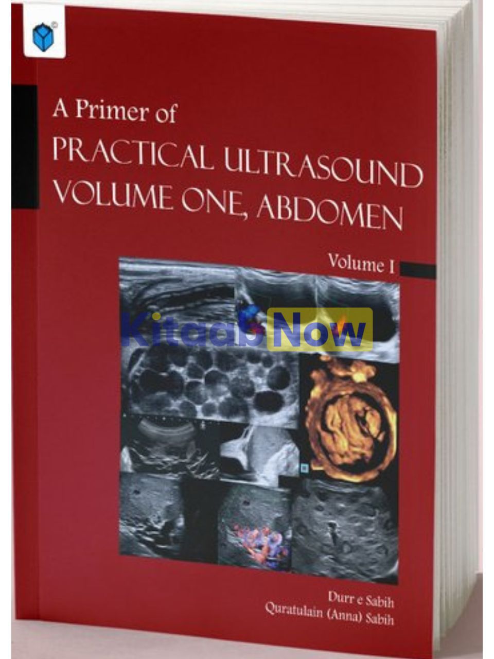
-22%
A Primer of Practical Ultrasound: Volume One, Abdomen (2nd Edition)
Generic
$ 7.42
Call 1800-313-2353 for expert assistance!
- Questions & Answers
DescriptionDr. Sabih has written a textbook that will capture the reader’s attention with high-quality images and a text that provides a scientific and enjoyable overview of the scanning techniques in the abdomen and pelvis. The text is easy yet fairly comprehensive and is sure to foster confidence in its users. As such, it deserves a place in every setting where diagnostic ultrasound imaging is used.PrefaceTo begin with, an apology; this second edition is unforgivably late. Although it is encouraging to see that trainee radiologists are still using photocopies of the first edition; our understanding of what we can see on ultrasound, how we can interpret ultrasound, and more importantly, how we can incorporate it in our clinical practice has changed tremendously since the last edition. A second edition with current knowledge was long overdue.Ultrasound was never meant to be within the sole purview of a single specialty. Developed by physicists and physicians with an interest in gynaecology and obstetrics, it has rapidly become indispensable for all who practice medicine. I struggle to think of a medical specialty where ultrasound is not used as a primary diagnostic tool or as a guide for other diagnostic or therapeutic procedures.It soon became evident that ultrasound needed to move out of the domain of the trained physician and could be used by nurses, emergency responders, and even battlefield medics to benefit even more patients.The field is on the cusp of yet another revolution, as machines with inbuilt artificial intelligence are poised to pass into the hands of non-trained staff in remote locations that are without appropriate technical expertise; and perhaps one day the patient herself. It teaches the user how to get the appropriate sections and then interprets the images. I know the Bill and Melinda Gates Foundation and others are already working on intelligent ultrasound machines that will not need a trained operator. This demonstrates that we have not yet exhausted the potential and promise of Ultrasound, and that is an exciting thought.However, I digress too much; back to this book!I have tried to write the book that I wish I had when I was learning ultrasound. The book starts by describing what is shown on the screen when the machine is first turned on, and then moves onto how the images are displayed, and then to how I actually do an abdominal ultrasound. I am particularly excited to include a chapter on “pearls of wisdom”, this chapter incorporates some of the tricks I have learned over the years that I find helpful with image interpretation; but there are hints and pointers peppered throughout that can make understanding the scan easier.The book is profusely illustrated, I have tried to include images that help clarify all of the concepts therein. I have used collages where possible to show a spectrum of variants of anatomy and changes in different stages of pathology. Line drawings are used for anatomical emphasis. Some of the images have been generously donated by friends from all over the world. I have acknowledged their contribution where these appear. The rest of the images are from my personal collection. Tables and text boxes highlight important points, list differential diagnosis and enumerate findings to help make the correct choice from the differential diagnoses. The clinical chapters are extensively referenced for the reader to peruse should he or she wish to learn more about a concept.I welcome my co-author, Dr. Quratul Ain (Anna) Sabih. Anna is a Consultant Surgeon at the Veterans Administration Medical Centre in Oklahoma City, USA. She insisted that we include clinical relevance instead of having a purely image interpretive text, and I agree that this has added value to the book. The clinical material will refresh memories and add new information enhancing rational patient management; obviously, which is the most important goal of our professional lives.This volume deals with abdominal ultrasound, the next volumes will deal with small parts, Doppler and Ob/Gyn, and will soon be available.I hope that this book will help the reader upskill and improve their confidence in using ultrasound to complement their clinical practice. Lastly, I wish the reader an accomplished professional career.– Durr-e-Sabih (Multan 2021)Table of ContentsChapter 01: Introduction to Ultrasound —————————————————————-1Chapter 02: Getting to Know Your Machine————————————————————6Chapter 03: Getting Started —————————————————————————-13Chapter 04: How I Do It ——————————————————————————–18Chapter 05: Pearls of Wisdom ————————————————————————-39Chapter 06: The Liver———————————————————————————–46Chapter 07: The Gallbladder and Bile Ducts———————————————————-134Chapter 08: The Kidneys, Ureters and Bladder ——————————————————-179Chapter 09: The Prostate and Seminal Vesicles ——————————————————270Chapter 10: The Spleen ——————————————————————————-280Chapter 11: The Pancreas —————————————————————————–293Chapter 12: The Adrenal Glands———————————————————————-304Chapter 13: The Lymph Nodes ———————————————————————–311Chapter 14: The Gastrointestinal Tract —————————————————————336Chapter 15: The Peritoneum, Peritoneal Cavity and Retroperitoneum —————————–373Chapter 16: The Anterior Abdominal Wall and Hernias ———————————————416Index—————————————————————————————————433ContributorsDr. Durr-e-Sabih established the first Department of Ultrasound at the Multan Institute of Nuclear Medicine and Radiotherapy in 1984. This was the first department of ultrasound for Nishtar Medical College and Hospital and all of South Punjab. He soon coined a new medical term for ultrasound diagnosis of hydatid cysts that is now accepted in literature. He established the first thalassaemia screening service for South Punjab and invented a device to help with chorionic villous sampling. A patent was granted to him for this. He proposed an Artificial Intelligence enabled computer software for analysis of liver texture and surface regularity using ultrasound. This paper is highly cited in the medical texture analysis literature. He proposed and created a prototype of an ultrasound training simulator.He is an IAEA expert and is Chair of the Asian Nuclear Medicine Board (ANMBoard.org).He was awarded an FRCPE in recognition of his contribution to Ultrasound and Nuclear Medicine by the Royal College of Physicians, Edinburgh. More recently, he was elected to a fellowship of the American Institute of Ultrasound in Medicine and awarded the much coveted FAIUM.He has published over 60 papers, 9 editorials, several book chapters and books. Some of the books by Dr. Durr-e-Sabih are given on the back cover.Dr. Quratulain “Anna” Sabih is a Clinical Assistant Professor of Surgery at the Oklahoma University Medical Centre and a Staff Surgeon at the Veterans Affairs Medical Centre in Oklahoma City, USA. Dr. Anna Sabih’s residency began in General Medicine. Very soon, she realised that surgery was her calling and she pursued a Surgical Residency at the University of New Mexico in Albuquerque, USA. Anna earned her Board Certification within weeks of completing her surgical residency and was elected as “TopDoc” by the Albuquerque Journal. She loves to teach and has received several best-teacher awards.Dr. Quratulain Sabih will be commencing a Breast Surgical Oncology fellowship at the Roswell Park Cancer Centre in Buffalo, New York. Dr. Quratulain Sabih has published several papers and book chapters.
Questions and answers of the customers
Only registered users are eligible to enter questions